HUMAN-MODEL-WORLD: A collection of scientific images
For the best immersive experience, we recommend to view the exhibition on a large screen.
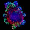
human embryo
Scientists use embryos to gain crucial insights into the development of genetic diseases, one of them being Huntington’s disease.
Huntington’s disease is a neurodegenerative disease that causes a breakdown of nerve cells in the human brain leading to problems with movement and cognitive impairments. The research group around Ali Brivanlou at The Rockefeller University uses alive cells of human embryos as well as synthetic embryos grown from stem cells to study Huntington’s disease. The mutation of a single gene kicks off the development of that disease which shows its first effects in these early human embryos.
Authors: Szilvia Galgoczi, Albert Ruzo, Christian Markopoulos, Anna Yoney, Tien Phan-Everson, Shu Li, Tomomi Haremaki, Jakob J. Metzger, Fred Etoc, and Ali H. Brivanlou
The Rockefeller University

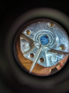
Authors: Claudio Bussi
The Francis Crick Institute

human lung cancer
Authors: David Novo, Zoe Ramsden, Camille Charoy, Charles Swanton and Erik Sahai
Credits: TRACERx Consortium
The Francis Crick Institute

Authors: Giulia Tyzack, Rickie Patani (UCL and the Francis Crick Institute), Jia Newcombe (NeuroResource)
The Francis Crick Institute

Author: Joaquina Delas Vives
The Francis Crick Institute

In order to fight disease, immune cells need to reach targets effectively, for this, they actively generate their own guiding system.
Part of a powerful immune response is the coordinated movement of immune cells during infection and inflammation, it is crucial that cells know where to go. But how do they do it? One type of immune cells, the so-called dendritic cells, plays a key role in this as they act like a messenger between the innate and adaptive response of the body: they scan tissues for intruders and migrate to the lymph nodes to relay information and initiate further responses. This movement is guided by chemokines, small signaling proteins released from the lymph nodes.
Authors: Jonna Alanko, Mehmet Can Ucar
Institute of Science and Technology Austria

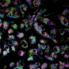
Unlocking the secrets of aging: Confocal microscopy reveals the complexity of senescent cell morphology, vesicle localization, and cytoskeletal rearrangements.
Cellular senescence, the biological phenomenon marking the irreversible cell cycle arrest associated with upregulated immune responses, plays a pivotal role in aging and age-related diseases. To understand the intricacies of cellular senescence, the Kono group at OIST employs advanced confocal microscopy. This technique allows us to investigate changes in senescent cell morphology and reveal the molecular mechanisms underlying these processes. These images contribute to a deeper understanding of the aging process and potential avenues for age-related diseases intervention.
Authors: Kamila Kozik
Okinawa Institute of Science and Technology

Tumour Family Trees
Cancer evolution through natural selection: Just as species adapt to their surroundings over time, tumours also evolve within the body, sometimes becoming resistant to treatments and spreading to other parts of the body.
When cancer cells divide via mitosis, they pass down their mutations to their daughter cells. As a result, cancer development can be modelled as an evolutionary process. To investigate patterns in tumour evolution, researchers in the Cancer Evolution and Genome Instability Laboratory at the Crick follow patients all the way from diagnosis through to either disease relapse or cure after surgery, tracking and analysing how their cancer develops. This image shows reconstructions of the tumour evolutionary trees of over 400 patients with early stage, untreated non-small-cell lung cancer.
Authors: Kristiana-Grigoriadis, Ariana Huebner, Abigail Bunkum, Emma Colliver, Alexander M. Frankell, Charles Swanton, Simone Zaccaria and Nicholas McGranahan
Credits: Mark S. Hill, Kerstin Thol, Mariam Jamal-Hanjani, CRUK, BMS, TRACERx consortium, TRACERx patients and their families
The Francis Crick Institute

Endothelial cells line the inside of blood vessels and regulate exchanges between the bloodstream and the surrounding tissues.
Authors: Michael Riedl
Institute of Science and Technology Austria

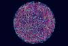
Wiseana Iridovirus
Authors: Nadishka Jayawardena
Okinawa Institute of Science and Technology

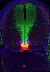
the cerebellum
Author: Nathaniel Heintz
Credits: Elitsa Stoyanova, Maria V. Moya
The Rockefeller University

The Paradox of Uniformity
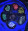
Can you spot the difference? When thousands of cancer cells are grown in parallel for use in research, scientists can interrogate the subtle differences across the seemingly identical 3D structures.
This image shows the three-dimensional growth of thousands of melanoma skin cancer cells for individual analysis. Cancer cells can be seeded in microwells - growing in parallel, one per microwell, in plates containing up to 20,000 microwells. In the absence of a surface to grow on to, the cancer cells will keep dividing and proliferating to form these spherical structures known as spheroids, that can then be used in experiments to mimic tumours.
Authors: Olivia Courbot, Alberto Elosegui-Artola
Credits: Alberto Elosegui-Artola, Brenda Canales, Maria Benito-Jardon
The Francis Crick Institute

Memory Neurons
Making proteins in brain cells visible can tell scientists a lot about their functions and how they affect memory formation and certain neurological diseases.
Neurons in your brain form an intricate web of connections. They continuously adapt these connections and create new ones. This way, they can form memories. Robert B. Darnell and his team from The Rockefeller University study the complex interplay of genes and proteins inside the brain that allows this to happen.
Authors: Caryn R. Hale, Kirsty Sawicka, Kevin Mora, John J. Fak, Jin Joo Kang, Paula Cutrim, Katarzyna Cialowicz, Thomas S. Carroll, and Robert B. Darnell
The Rockefeller University

Authors: Simon Butterworth
The Francis Crick Institute

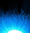
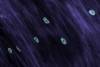
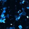
Scientists study the intricate self-regulation and repair mechanisms of DNA to understand tumor formation and the process of aging.
Your DNA is an incredibly long and thin molecule that is wound up in packages called chromosomes in each of your body’s cells. The ends of the DNA strands are protected by a special section of DNA called telomere which shortens over time as you age. Titia de Lange and her team at The Rockefeller University study how this shortening helps the body to suppress tumors from growing and how it interacts with DNA repair mechanisms. The image shows human chromosomes in blue with their telomeres in green.
Authors: Isabelle Schmutz, Arjen R. Mensenkamp, Kaori K. Takai, Maaike Haadsma, Liesbeth Spruijt, Richarda M. de Voer, Seunga Sara Choo, Franziska K. Lorbeer, Emma J. van Grinsven, Dirk Hockemeyer, Marjolijn C.J. Jongmans, and Titia de Lange
The Rockefeller University

Authors: Tony Fearns
The Francis Crick Institute

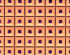
The human body is a complex system composed of multiple organs, each intricately organized from billions of cells working in unison as specialized units to sustain our health and well-being. This meticulous cellular architecture is essential for orchestrating interconnected processes. Importantly, in the face of disease, it is at this cellular level that the primary battleground emerges.
In the laboratory, doctors and scientists use cell cultures as indispensable tools for studying and developing treatments for diseases. By cultivating cells in controlled environments outside the body, researchers can examine cellular behavior and responses to various conditions.
The NEURAD technology developed at OIST allows scientists to guide the growth and division of cultured cells along predetermined patterns.
Authors: Zakaria Ziadi, Dimitar Dimitrov
Credits: NEURAD
Okinawa Institute of Science and Technology
sensory structures
Authors: A. James Hudspeth, Agnik Dasgupta, Caleb C. Reagor, Sang P. Paik, Lauren M. Snow, Adrian Jacobo
The Rockefeller University

Authors: Alessandro Alberto
Okinawa Institute of Science and Technology

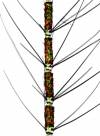
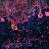
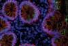
Did you know there’s a ‘second brain’ in the gut, with around 100 million nerve cells, more than in the spinal cord?
In this image, scientists have used fluorescent labelling to capture the beauty of the ‘second brain’, known as the enteric nervous system. This ‘brainbow’ is made of fluorescently labelled neurons in the adult mouse gut, where each colour labels an individual type of neuron and its projections, giving each a unique signature. The connections between them, the ganglia, can be found at the junction of these tracts. Collectively this network of cells – the enteric nervous system – independently orchestrate various gastrointestinal functions, such as sensing changes in the environment and coordinating muscle movements and gland secretions.
Autors: Alvaro Castano Medina, Yuuki Obata
The Francis Crick Institute

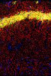
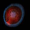
The code that the brain uses to communicate about smell originates in the nose.
The image shows a section of a mouse’s nasal cavity imaged with a confocal microscope. The air the animal breathes passes through this convoluted structure, lined with olfactory sensory neurons. This is where odor molecules are first detected and converted into electrical signals used by the brain. Each sensory neuron is a specialist: it chooses to express one type of detector out of hundreds of detector types. As a result, each one responds to just a handful of odorous molecules.
Authors: Cary Zhang, Izumi Fukunaga
Credits: Imaging section, OIST Graduate University
Okinawa Institute of Science and Technology

Authors: Colin Ratcliffe, Erik Sahai
The Francis Crick Institute

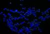
Gut Lymph Node
Studying gut lymph nodes tells researchers about how the immune system manages the myriad of microbes living in the intestines.
The gut is a turbulent place, where hundreds of species of bacteria live alongside whatever microbes happen to have hitched a ride in on your lunch. Daniel Mucida and his team at The Rockefeller University investigate which microbes are allowed to thrive and whose spread is culled in a delicate trade-off by the immune system and how this affects our health. The image shows the gut lymph node of a mouse which the scientist study to find out how they influence this balance.
Authors: Carla R. Nowosad, Luka Mesin, Tiago B. R. Castro, Christopher Wichmann, Gregory P. Donaldson, Tatsuya Araki, Ariën Schiepers, Ainsley A. K. Lockhart, Angelina M. Bilate, Daniel Mucida, and Gabriel D. Victora
The Rockefeller University

Juvenile Frog Leg
Authors: David Vijatovic
Institute of Science and Technology Austria

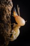
Studying social insects, who operate as a singular cooperative unit to fight disease, can teach us many aspects about the workings of immune systems, even our own.
To reduce the risk of infection and disease transmission throughout their colony, ants utilize various methods ranging from collective nest hygiene, isolation to mutual sanitary care. At ISTA, Florian Strahodinsky and his colleagues of the Cremer Group study the collective protection mechanisms that emerge from social interactions and individual behaviors of the ants.
Authors: Florian Strahodinsky
Institute of Science and Technology Austria


Authors: Gayathri Singaraju, Robert Hauschild
Institute of Science and Technology Austria

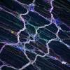
Immune Cells migrating over a nanofabricated structures that triggers an actin accumulation, observable as bright stripes.
Authors: Isabelle Mayer, credit to Florian Gaertner for piloting the project
Institute of Science and Technology Austria

Authors: Leslie B.Vosshall
Credit: Margaret Herre
The Rockefeller University

within a fruit-fly testis
Authors: Li Zhao, Evan Witt, Sigi Benjamin, Nicolas Svetec
The Rockefeller University

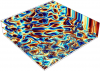
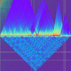
Massive numerical simulations show how two-dimensional turbulence transitions to three-dimensional when in proximity to a rigid wall.
Despite their size, large-scale flows in the atmosphere act like two-dimensional turbulence due to the horizontal lengths being much larger than the height of the atmosphere. Two-dimensional turbulence is characterized, by a portion of the energy flowing towards the large eddies, the swirls and their reverse currents in a fluid, which deviate from the general direction of the fluid. This is in contrast to conventional turbulence in three-dimensions in which any injected energy is transferred from large to small eddies.
This image shows that away from the wall, at the top, turbulence is two-dimensional and characterized by coherent and large vortices. On the other hand, at the bottom the wall causes turbulence to become three-dimensional, characterized by the presence of small-scale vortices.
Authors: Marco Rosti
Okinawa Institute of Science and Technology

Authors: Mary E. Hatten, Hourinaz Behesti, Taylor R. Fore, Peter Wu, Zachi Horn, Mary Leppert, Court Hull
The Rockefeller University

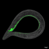
Its transparent body and consistent cell number make C. elegans a great model organism to study a wide range of biological phenomena.
Authors: Michaela Mišová, Chris Li
Institute of Science and Technology Austria

Authors: Rashmi Priya, Srinivas Allanki
The Francis Crick Institute

Authors: Silvia Jamrichova
Institute of Science and Technology Austria

Authors: Stephane Mouilleron
Credit: Martina Wirth
The Francis Crick Institute

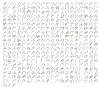
Mapping how DNA folds in space: Compressing four metres of DNA into the tiny nucleus of a cell is equivalent to fitting 40 km of very fine thread inside a tennis ball – and that requires some very organised packing!
These long strands of DNA must fit inside a tiny cell nucleus, two million times smaller than the length of the genome. Rather than being randomly folded, the DNA is carefully organised into defined loops and coils by groups of specialised proteins. This structured way of condensing the DNA prevents tangling and allows it to become available to the many molecules that replicate it, repair it and access its genes.
These maps show, at an unprecedented resolution, how this folding works in yeast (Saccharomyces cerevisiae). Warmer colours show connections between sections of DNA which loop near to each other, while colder areas are largely devoid of encounters.
Authors: Thomas M. Guérin, Christopher Barrington, Frank Uhlmann
Credits: Software used to display the genomic interaction data: Kerpedjiev et al. (2018) HiGlass: Web-based visual comparison and exploration of genome interaction maps. Genome Biology, 19:125.
The Francis Crick Institute

Pure mathematics sometimes asks seemingly simple questions and finds complex answers addressing decades-old ideas and conjectures.
Research in pure mathematics can seem strange to the uninitiated but understanding the hidden relations between numbers and many other mathematical objects can reveal new avenues to understanding the world. Tim Browning, professor at ISTA, studies a branch of mathematics called analytical number theory and delves deep into the foundations of the field.
Authors: Régis de la Bretèche, Tim Browning, Emmanuel Peyre
Institute of Science and Technology Austria

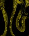
Shining a light on mini-guts: Human cells grown in a dish in the lab are crucial research tools that allow scientists to model development and diseases.
Organoids are one of the recent biomedical research breakthroughs that allow scientists to investigate how human organs grow and respond to medical treatments. This is a fluorescent image of an organoid, or ‘mini-gut’, grown in a petri dish in the lab. They are essentially stem cells growing three dimensionally to form a little cluster of cells replicating the structure and function of a specific organ, in this case the small intestine. Understanding how these cells function in healthy guts and what happens when tumours arise helps scientists to develop specific drugs for bowel cancer treatment. In addition, they can use this knowledge to grow replacement human gut tissue in the lab to use in organ transplants or for testing new drugs.
Authors: Vivian Li
The Francis Crick Institute

Authors: Yvonne Vallis, Saliha Ece Sönmez
Institute of Science and Technology Austria

Authors: Ales Bucek
Okinawa Institute of Science and Technology

Authors: Alessandro Monti
Okinawa Institute of Science and Technology

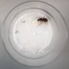
Although this image is not from research project per se, it still shows us how exciting and informative a scientific view on to everyday occurrences can be:
The 3-4 layers and the resulting size of the hailstone show that the grain probably rose and fell several times in a cumulonimbus cloud before it finally fell to earth - a phenomenon that is becoming increasingly common due to climate change.
Authors: Bidyut Goswami
Institute of Science and Technology Austria

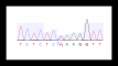
kill her children
A genetic mystery and miscarriage of justice: Kathleen Folbigg was found guilty of killing her four children, but science revealed that their sudden deaths could be explained by genetic conditions.
For years, Carola Vinuesa and her team have been studying the mutations linked to rare autoimmune diseases, including Lupus, with a view to guiding precision treatments. But she was enlisted to investigate a complex case where genetic mutations may have been the culprit in a tragic murder conviction. This image illustrates the genetic finding key to the release and exoneration of Kathleen Folbigg, a mother who spent 20 years in prison after being wrongly convicted of murdering her four children. The central wave of the DNA sequencing analysis shows a key variation (C/T) in a gene called CALM2, shared by Kathleen and her two daughters. In the 2023 legal inquiry into Kathleen’s convictions, a cardiac arrest caused by this variant was accepted as a reasonable explanation for the sudden deaths of her daughters. The Folbigg sons also suffered from respiratory problems and severe epilepsy. This was the first time in the world that genomic medicine was used in a courtroom to identify a cause of death.
Authors: Carola G. Vinuesa, Todor Arsov, Vicki Athanasopoulos, Yafei Zhang
The Francis Crick Institute

porous media
Revealing the unseen currents of life: OIST researchers illuminate the hidden dynamics of fluids as they wander through tiny porous landscapes, mirroring the essential flow within our bodies and the lifelines of our urban fabric.
In the quest to understand the hidden flows that are applicable from biology to urban life, the Micro/Bio/Nanofluidics unit at OIST ventures into the microscopic world. Here, they trace the journey of fluids as they navigate through a maze of glass beads, each as thin as a hair. This delicate exploration, using X-rays combined with flow field measurements, unveils a three-dimensional dance of fluids within these minuscule spaces.
Authors: Daniel Carlson, Kazumi Toda-Peters, Amy Q. Shen, Simon J. Haward
Okinawa Institute of Science and Technology

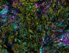
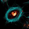
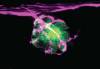
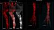
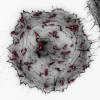
A viral takeover: Vaccinia virus hijacks the host cell’s actin cytoskeleton (grey) to reach the surface, where it creates protrusions called ‘actin tails’ (red) to project into neighbouring uninfected cells.
Vaccinia virus is a poxvirus similar to smallpox, but less harmful. The virus hijacks many systems in human cells to stimulate its replication and spread. This includes the actin cytoskeleton (grey), which can be thought of as scaffolding that maintains the shape and structure of cells and also helps them move. The virus uses the filaments of the cytoskeleton to reach the surface of the cell to generate ‘actin tails’ (red). These small protrusions extend outside of the cell, helping propel the virus towards neighbouring cells to enhance the spread of infection. By staining actin with a fluorescent red dye, and imaging the infected cell in a microscope, it was possible to see how the virus manipulates and changes human cells for its own benefit.
Authors: Gabrielle Larocque
Credits: Michael Way, Gruppenleiter, Cellular Signalling and Cytoskeletal Function Laboratory,
The Francis Crick Institute

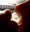
Not all that is commonly known, is fully understood: At ISTA, researchers study electrostatic in all its intricacies.
In this setup, with the help of a small glass sphere bouncing on a plate, one can measure electric charge incredibly precise manner as charge exchanges at every “dribble”, aka touch of the surface.
Authors: Galien Grosjean
Institute of Science and Technology Austria

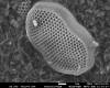
of Discochiton
Authors: Jinyeong Choi
Okinawa Institute of Science and Technology

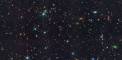
From here to the edge of the universe: Photos taken with the James Webb Space Telescope are helping researchers understand how galaxies and black holes formed during the earliest stages of the universe.
Galaxies such as our own Milky Way are the largest bound structures in our Universe and consist of gas, stars, planets, black holes and dark matter. The Matthee group at ISTA investigates the physical mechanisms that determine how galaxies and their components form and evolve, using observations of the very distant universe. For this, they take pictures of faraway segments of the universe and analyze them almost pixel by pixel: Each bit of information about color, gradients, shape, blurriness or the relative positions of objects to each other.
Authors: Jorryt Matthee, Daichi Kashino (National Astronomical Observatory of Japan), Ruari Mackenzie (ETH Zurich), Simon Lilly (ETH Zurich), NASA, ESA, CSA
Institute of Science and Technology Austria

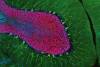
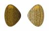
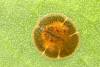
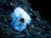
anemone fish
The remote Ogasawara Islands, only reachable via a 24 hours boat ride from Japan, contain unique ecosystems and host many endemic species and a very special population of the anemone fish Amphiprion clarkii.
While adult anemone fish are usually orange with white bands, adults in this population are black with white bands. The individual in the picture is a juvenile, it is still yellow and will change to black as it grows. During a sampling trip to study the ecology and genetics of these fish, researchers noticed that many of the host sea anemones, as well as some corals, were bleached. Bleaching occurs when rising water temperatures cause the anemone to lose its symbiotic algae. Bleaching has a negative effect on the growth of the anemone and can even lead to its death. Ultimately, the anemone fish will die as well, as they cannot survive without their host. The consequences of climate change do not spare any ecosystem, even in the most remote places.
Authors: Manon Mercader
Okinawa Institute of Science and Technology

Authors: Marco Rosti
Okinawa Institute of Science and Technology

Authors: Mehmet Arif Zoral
Okinawa Institute of Science and Technology

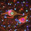
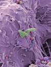
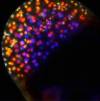
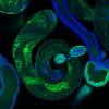
Immune Response
Researchers study how the immune system keeps adapting its response to intruders like SARS-CoV-2 even months after being sick.
The human body has to defend itself against a wide variety of intruders ranging from microbes to viruses. The adaptive part of the immune system detects them and then produces specific receptors to recognize the intruders if they should appear again. Over time, the immune system even refines these receptors to become more effective. For one of their studies, Michel C. Nussenzweig and his colleagues produced the image above showing SARS-CoV-2 (green) in intestinal cells three months after infection in order to investigate how the body continues to adapt its immune response to it.
Authors: Christian Gaebler, Zijun Wang, Julio C. C. Lorenzi, Frauke Muecksch, Shlomo Finkin, Minami Tokuyama, Alice Cho, Mila Jankovic, Dennis Schaefer-Babajew, Thiago Y. Oliveira, Melissa Cipolla, Charlotte Viant, Christopher O. Barnes, Yaron Bram, Gaëlle Breton, Thomas Hägglöf, Pilar Mendoza, Arlene Hurley, Martina Turroja, Kristie Gordon, Katrina G. Millard, Victor Ramos, Fabian Schmidt, Yiska Weisblum, Divya Jha, Michael Tankelevich, Gustavo Martinez-Delgado, Jim Yee, Roshni Patel, Juan Dizon, Cecille Unson-O’Brien, Irina Shimeliovich, Davide F. Robbiani, Zhen Zhao, Anna Gazumyan, Robert E. Schwartz, Theodora Hatziioannou, Pamela J. Bjorkman, Saurabh Mehandru, Paul D. Bieniasz, Marina Caskey, and Michel C. Nussenzweig
The Rockefeller University

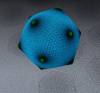
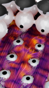
Particles of Light
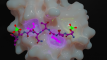
Light is around us all the time — in visible or invisible form — and scientists can use it to explore the quantum-mechanical nature of our reality.
Experimental physicists employ devices made from special kinds of glass and other materials like the one shown in this image to manipulate light and to achieve quantum-mechanical effects that are impossible in our everyday world.
Authors: Rishab Sahu, Thomas Werner, Liu Qiu
Institute of Science and Technology Austria

Authors: Saacnicteh Toledo Patino
Okinawa Institute of Science and Technology

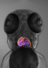
Animal behavior is based both on instinct and learning. Researcher study animal brains down to the individual neurons to untangle these complex influences.
Even the tiniest insects like fruit flies can learn something new—they must to survive. Vanessa Ruta and her team study which animal behaviors are predetermined by their genes, which ones are learned from experience, and how these two interact. For this, they look at the relatively simple and very well understood neural circuitry of fruit flies. The image shows specific neurons in a fruit fly’s ‘brain’ that take in and process signals from smelling the environment letting the animal learn what they mean and influencing its behavior.
Authors: Vanessa Ruta, Annie Handler. Thomas G.W. Graham, Raphael Cohn, Ianessa Morantte, Andrew F.Siliciano, Jianzhi Zeng, Yulong Li
The Rockefeller University